
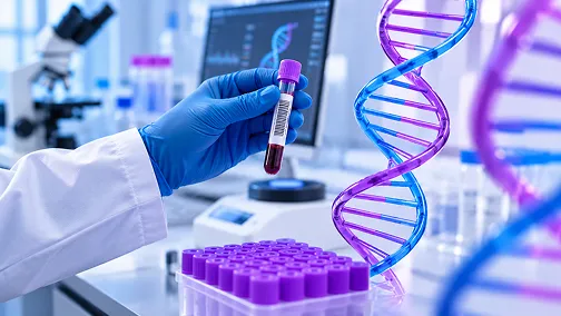
For the patient, a genetic test usually feels simple: a short conversation, a small blood sample, and a report a few days or weeks later. Behind that report lies a careful journey through the laboratory, combining chemistry, powerful machines, computers and expert human judgement. This article follows a sample from the clinic to the final diagnosis.
It begins with a conversation
Every good genetic test starts before the sample is taken. The doctor or genetic counsellor talks through the patient's symptoms and family history, chooses the test that best fits the question, and explains what the test can and cannot show. The patient then gives written consent, including a choice about whether to learn of any unexpected findings unrelated to the reason for testing.
The sample
Most tests need only 2 to 5 ml of blood collected in a tube containing an anticoagulant called EDTA. Saliva or a cheek swab can sometimes be used instead. In pregnancy, cells from the placenta are collected by chorionic villus sampling (CVS), usually between 11 and 14 weeks, or fluid around the baby is collected by amniocentesis, usually from 15 weeks onwards. After a miscarriage, a small piece of pregnancy tissue can be tested. For cancer, the laboratory usually works with tumour tissue already removed during a biopsy or surgery. Proper labelling and safe transport at the right temperature matter as much as anything that follows.
Inside the laboratory
The first step is to extract pure DNA from the cells and check that there is enough of it, and that it is of good quality. What happens next depends on the test chosen.
For karyotyping, living cells are grown in culture for several days, then their chromosomes are stained and examined under a microscope. This shows extra or missing chromosomes and large rearrangements, including balanced translocations, but it cannot see changes smaller than about 5 to 10 million DNA letters. Chromosomal microarray works directly on DNA, without growing cells, and detects missing or extra pieces about 100 times smaller than karyotyping can see. It cannot, however, detect balanced rearrangements. For a single known change, simpler methods such as PCR and Sanger sequencing read just that small region.
Larger tests use next-generation sequencing (NGS). The DNA is broken into millions of short fragments and tagged so that each can be tracked. For an exome or gene panel, the fragments of interest are first fished out from the rest. The sequencing machine then reads all of them at the same time, and each position in the DNA is read many times over so that occasional reading errors can be spotted and ignored.
From raw data to meaningful results
A single exome produces billions of letters of raw data, so the next stage belongs to computers. Specialised software lines up each fragment against the standard human reference genome, much like fitting pieces of a jigsaw puzzle, and lists every place where the patient's DNA differs. This usually produces tens of thousands of variants.
The software then adds information about each variant: how common it is in healthy people, including Indian reference data from the GenomeIndia Project, whether it has been reported before in patients, and how it is likely to affect the gene. Common harmless variants are filtered out, and the remaining few are matched against the patient's symptoms.
The human touch: interpretation
Computers can narrow the list, but people make the final call. Trained scientists and clinical geneticists review each remaining variant using international guidelines from the American College of Medical Genetics and Genomics (ACMG) and classify it as pathogenic, likely pathogenic, of uncertain significance, likely benign or benign. They check whether the variant fits the patient's symptoms and the way the condition is inherited. Important findings may be confirmed with a second method, and testing the parents can show whether a variant was inherited or is new in the child.
The report and beyond
The final report explains the main finding, its classification, the condition it is linked to, how it is inherited and what it may mean for the family. It also states the limits of the test. A targeted test may take a few days; exome or genome sequencing usually takes a few weeks.
Quality matters at every stage. In India, laboratories accredited by NABL to the international ISO 15189 standard follow strict procedures and take part in external quality checks, which helps ensure that results are accurate and reliable.
The journey does not end with the report. The doctor or genetic counsellor explains the result, discusses next steps in treatment or monitoring, and, where relevant, offers testing to other family members. Families can help the whole process by sharing complete medical records, describing symptoms clearly and making samples from both parents available when asked.